Packages

Demo Modules

Our demo package contains a variety of example training modules which offer immediate feedback on diagnostic accuracy.

Nasogastric Tube Position on Chest X-ray
 Feeding a patient through a misplaced nasogastric tube is a "never-event". Developed in collaboration with BSGAR, RCR, SOR and BAPEN, this package provides real-world cases, supported by step-by-step teaching, to help you train to check the positioning of nasogastric tubes on chest radiographs and confirm that they are safe to use.

Chest X-ray Fundamentals
 Chest X-rays are the most frequently requested imaging investigation worldwide. A variety of health care professionals including doctors, nurses and radiographers are required to review chest X-rays. Curated by expert thoracic radiologists, this package aims to provide an introduction to chest X-ray interpretation.

Emergency Radiology

Equip yourself for working out of hours and for radiology emergencies with our modules developed in collaboration with the London School of Clinical Radiology. This package includes ultrasound, X-ray, CT and MRI cases with detailed explanations covering acute abdominal, neurological, spinal, paediatric, thoracic, vascular and trauma imaging.

Acute Pathology on Head CT
 A head CT is the most common referral of cross-sectional imaging made by emergency departments. This package provides teaching and training using unenhanced CT images with examples of common abnormalities such as ischaemia, haemorrhage and space occupying lesions.
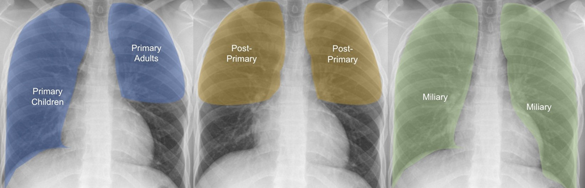
Tuberculosis on Chest X-ray
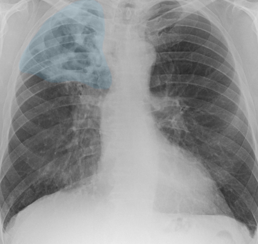
Tuberculosis (TB) is a highly infectious disease caused by bacteria, and its diagnosis relies on chest X-rays. This package helps explain the pathogenesis of TB, its appearance on radiographs and offers guidance on features to consider that differentiate latent and active TB infection

Prostate mpMRI Training
 Developed in collaboration with Prostate Cancer UK and expert uroradiologists, this package provides content focused on the interpretation and reporting of prostate multiparametric MRI scans.
There are several modules, each with 25 cases, that form a comprehensive package of training material enabling you to improve mpMRI diagnosis skills.

Breast Imaging Training
 A collaboration between the National Breast Imaging Academy (NBIA) and RAIQC with a range of teaching case modules to support those training in breast imaging.
This package provides a valuable resource for radiology trainees, particularly those sitting the FRCR 2B, as well as breast clinicians and advanced practitioner/consultant radiographers.

Lines, Tubes and Devices on Chest X-ray
 Rapid recognition of incorrect placement of devices, lines, or tubes on chest radiographs is a key skill and vital to ensuring patient safety. This package covers the basics of airway management devices, vascular lines, pleural drains, cardiac conduction devices and nasogastric tubes seen on chest X-rays.

Renal Transplant on Ultrasound
![]()
This package introduces you to the assessment of transplant kidneys using ultrasound.
You will learn the basics of ultrasound physics and imaging, an approach to scanning the kidneys and the appearances of both normal transplant kidneys and pathology.
Pulmonary Nodules on CT
![]()
A chest CT scan is a critical tool in the assessment of pulmonary nodules. Based on the British Thoracic Society guidelines, this package includes concise educational materials explaining the assessment of lung nodules, as well as real-world training cases to develop your CT interpretation skills.

Interstitial Lung Disease on HRCT
![]()
High-resolution CT (HCRT) is a key diagnostic tool for assessing the extent of Interstitial Lung Disease and exploring possible aetiologies. This package will teach you the manifestations of ILD on HRCT and train you in the fundamentals of interpretation.

COVID-19 on Chest CT and X-ray
 Our COVID-19 content is for healthcare professionals wishing to accurately diagnose COVID-19 on chest X-ray and CT scans. It trains users to identify the presence and severity of the infection, and to differentiate these from other conditions that may present in a similar manner.