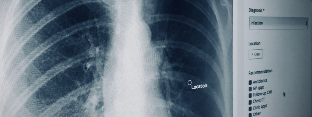
Chest X-rays are non-invasive, low cost, and provide important information regarding the anatomy and potential pathology of the structures of the chest, such as the lungs, mediastinum and rib cage. Because of this, the chest X-ray is the most frequently requested imaging investigation worldwide. They are often reviewed and acted upon by many different healthcare professionals, including doctors, nurses and radiographers.
In October 2021, the Healthcare Safety Investigation Branch (HSIB) published an independent report titled Missed detection of lung cancer on chest X-rays of patients being seen in primary care1, making the safety observation:
It may be beneficial if existing educational platforms were used to support healthcare staff who report on chest X-rays with their ongoing professional development and demonstration of the clinical quality of their work
In the same report, RAIQC is given as an example of such a tool designed to both develop and enable self-assessment of skills in interpreting chest X-rays.
This Chest X-rays Fundamentals package has been developed by expert thoracic radiologists to clearly convey the principles of chest X-ray interpretation, demonstrate how common and important abnormalities are identified, and describe how these are usually managed. The educational content provides a foundation of knowledge which is then applied in interpreting actual clinical cases. Training cases enable the development of fundamental interpretation skills with immediate feedback and assessment cases allow the newly acquired skills to be demonstrated.
The pathologies explored in this package are:
- Pneumothorax
- Pleural effusion
- Pneumonia
- Lung collapse
- Malignancy
- Hilar lymphadenopathy
- Heart failure
- Interstitial lung disease
- Pneumoperitoneum
- Foreign bodies
Upon completion of the available modules, reporting statistics are calculated, and certificates can be downloaded or shared so as to show your continuous professional development.
Key benefits:
- Obtain vital understanding of the manifestations of common and important thoracic pathologies on chest X-ray
- Build your interpretation skills with a diverse list of real-world cases
- Test your ability in a safe, simulated environment
- Earn CPD points at your own pace
References
- Healthcare Safety Investigation Branch (October 2021) Missed detection of lung cancer on chest X-rays of patients being seen in primary care
This package includes the following modules:
-
2
Chest X-ray - Educational content
Teaching material with real-world cases explaining how to identify common and important pathologies on CXR.
-
2
50
Chest X-ray Fundamentals - Training cases
A series a Chest X-rays for you to practise spotting important pathologies on CXR, with immediate feedback provided on your accuracy.
-
1
30
2h
Chest X-ray Fundamentals - Assessment cases
Test how well you can interpret CXRs, with a summary of your accuracy provided on module completion.
You must sign in or register to access this package
Package Summary
- Chest X-ray Fundamentals
- Created 21 April, 2023
-
Module types included:
- Educational content 1
- Training 1
- Assessment 1
- Training and assessment cases 80
- CPD points available 5
